News Category
- Blog Announcements

Dr. Susan Gauthier’s research passion lies in uncovering the underlying principles that govern disease progression in Multiple Sclerosis (MS). Despite potent anti-inflammatory treatment and other strides in MS, predicting a patient’s disease progression still eludes us. Furthermore, there has been very little impact on altering the clinical course of MS once patients enter the progressive phase.
Her research focuses on exploring two main hypotheses on why people may progress:
- Years of loss of myelin (demyelination) leave axons vulnerable to degeneration (or dying off).
- Chronic activation of immune cells (microglia) within the brain may be toxic to neurons.
The research tools her team uses to investigate these theories include MRI-based myelin water fraction imaging, which allows us to quantify myelin in white matter and follow it over time, and Positron Emission Tomography (PET) to label microglia or the immune cells in the brain.
In addition, Dr. Gauthier, in collaboration with Weill Cornell Medicine physicist Yi Wang, Ph.D., has utilized a novel MRI technique called Quantitative Susceptibility Imaging (QSI) to explore early iron deposition and chronic MS lesions.
Findings from her research exploring MS lesions through these imaging tools have led to a number of publications. Published in Frontiers of Neurology, Dr. Gauthier’s team demonstrated that the relationship of the loss of myelin in lesions and normal brain tissue is associated with a loss of neurons. They also demonstrated that myelin recovery does occur in early MS lesions (published in Neuroimage Clinical). The team is now exploring potential repair and ongoing damage in chronic (or older) MS lesions.
They have demonstrated that those chronic lesions that have a rim of iron have more myelin damage (published in American Journal of Neuroradiology) and are more likely to have more chronic inflammation as measured on PET (presented at ECTRIMS 2017). This has lead to an appreciation that chronic lesions are not just scars, may continue to cause damage and should be a target for new treatments.
Dr. Gauthier’s strategy is to combine imaging tools to dissect lesions to acquire information which can be utilized to develop novel treatment targets for both re-myelination and myelin protection. The ultimate goal of her research is to identify potential mechanisms that will decrease the amount of myelin damage and/or stimulate myelin repair within lesions.
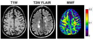
Example of conventional MRI demonstrating MS lesions and Myelin Water Fraction (MWF) - this shows lower myelin in MS lesions
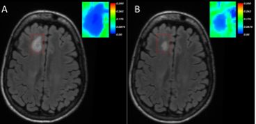
Example of a new lesion in panel A and MWF of a lesion (inset) - improvement of a lesion and MWF is shown in panel B
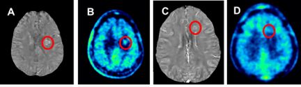
Example of a rim of iron in lesions on QSM demonstrating higher inflammation on PET scans.